


Enfermera jubilada recupera la vista después de extirparle un tumor cerebral
PENSACOLA, Fla. – Cuando Kathy Stinson comenzó a experimentar cambios en la visión, no estaba muy preocupada porque había usado lentes correctores desde que tenía 7 años.
“Pensé que solo necesitaba una actualización de la receta,” explicó Stinson, de 61 años. “Pero después de que mi optometrista aumentó mi receta, la visión en mi ojo izquierdo todavía estaba borrosa. Entonces empecé a tener problemas para leer, y manejar se volvió más difícil.”
Stinson regresó al optometrista un par de meses después. Su pérdida de visión progresiva marcó un problema que iba más allá de un cambio de receta. Esta vez su optometrista la instó en hacer un seguimiento con su médico de atención primaria. Ordenó una resonancia magnética, que reveló un tumor del tamaño de una naranja que presionaba el lóbulo frontal de su cerebro.
La visión borrosa de Stinson fue causada por un meningioma, un tumor que se forma en las membranas que cubren el cerebro. El tumor presionaba su corteza frontal, la parte del cerebro que controla el pensamiento y la toma de decisiones. También controla la visión. La mayoría de los meningiomas crecen muy lentamente, a menudo durante muchos años sin causar síntomas. Cuando aparecen los síntomas por primera vez, suelen ser dolores de cabeza y convulsiones, causados por el aumento de la presión del tumor en crecimiento.
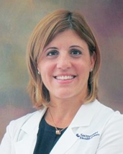
Debido al tamaño y la complejidad del tumor, Stinson fue remitida a la doctora María Toledo, una neurocirujana en el Hospital Sacred Heart de Pensacola, que realiza este tipo de cirugía compleja.
“El tumor era tan grande que estaba envuelto alrededor de los principales vasos sanguíneos en su lóbulo frontal,” dijo Toledo. “Cuando un tumor se ha abierto camino en el cerebro, es un delicado equilibrio extraerlo sin dañar las áreas circundantes.”
Antes de la cirugía, Toledo utilizó imágenes por resonancia magnética y tomografía computarizada para determinar la ubicación exacta y el tamaño del tumor. Los escáneres cerebrales también revelaron una protuberancia similar a un globo (aneurisma) que podría causar un accidente cerebrovascular si no se trata. Durante el procedimiento de 10 horas, Toledo extrajo el 99 por ciento del tumor.
Dos semanas después de su cirugía cerebral en marzo, Stinson regresó al hospital para reparar el aneurisma. Stinson, quien es una enfermera ortopédica retirada, dijo que le dio tranquilidad tener a Toledo en la sala de operaciones.
“La neurocirugía es algo más que establecer un yeso en un hueso roto; Estoy agradecida de tener a alguien con la experiencia de Dra. Toledo para que realice mi cirugía,” dijo. “Ella ya ha salvado mi vida dos veces. Si no fuera por ella, no estaría aquí ahora.”
Lentamente, Stinson está recuperando la visión en su ojo izquierdo. Ella espera poder conducir de nuevo.
La Dra. María Toledo obtuvo su título de médico en la Universidad de Puerto Rico y completó su entrenamiento de residencia en neurocirugía en la Escuela de Medicina de Puerto Rico.
Retired nurse regaining eyesight after brain tumor is removed
 PENSACOLA, Fla. — When Kathy Stinson started experiencing vision changes, she wasn’t too concerned because she had used corrective lenses since she was 7.
“I thought I just needed a prescription upgrade,” Stinson, 61, explained. “But after my optometrist increased my prescription, the vision in my left eye was still blurry. Then I started having trouble reading and driving became more difficult.”
Stinson returned to the optometrist a couple months later. Her progressive vision loss signaled a problem that went beyond a prescription change. This time her optometrist urged her to follow up with her primary care doctor. He ordered an MRI, which revealed an orange-sized tumor that was pressing on her brain’s frontal lobe.
Stinson’s blurred vision was caused by a meningioma, a tumor that forms on membranes that cover the brain. The tumor was pressing on her frontal cortex, the part of the brain that controls thinking and making decisions. It also controls vision. Most meningiomas grow very slowly, often over many years without causing symptoms. When symptoms first appear, they are often headaches and seizures, caused by increased pressure of the growing tumor.
Because of the tumor size and complexity, Stinson was referred to Dr. Maria Toledo, a neurosurgeon at Sacred Heart Hospital Pensacola, who performs this kind of complex surgery.
“The tumor was so large that it was wrapped around major blood vessels in her frontal lobe,” Toledo said. “When a tumor has worked its way deep into the brain, it’s a delicate balance to extract it without damaging surrounding areas.”
Before surgery, Toledo used MRI and CT scans to map out the exact location and size of the tumor. The brain scans also revealed a balloon-like bulge (aneurysm) that could cause a stroke if it was left untreated. During the 10-hour procedure, Toledo removed 99 percent of the tumor.
Two weeks after her brain surgery in March, Stinson returned to the hospital to repair the aneurysm. Stinson, who is a retired orthopedic nurse, said it gave her peace of mind to have Toledo in the operating room.
“Neurosurgery is more than just setting a cast on a broken bone; I’m thankful that I had someone with Dr. Toledo’s expertise perform my surgery,” she said. “She’s saved my life twice already. If it wasn’t for her, I wouldn’t be here now.”
Slowly, Stinson is regaining the vision in her left eye. She looks forward to being able to drive again.
Dr. Maria Toledo earned her medical degree at the University of Puerto Rico and completed her residency training in neurosurgery at the Puerto Rico School of Medicine.


